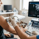
The years Bernhard Scheja spent practising in Switzerland gave him an unusually thorough grounding in emergency sonography — a discipline where speed, accuracy, and clinical confidence must work in perfect alignment.
In acute medical settings, the margin for diagnostic error is narrow. Patients presenting with chest pain, severe abdominal discomfort, or haemodynamic instability require rapid, reliable assessment — and the clinician who can perform and interpret an ultrasound scan at the bedside holds a significant advantage. Doctor Bernhard Scheja developed much of his emergency sonography expertise during his time in Switzerland, where demanding clinical environments and rigorous institutional standards shaped a practice that remains defined by speed without compromise on accuracy.
Emergency sonography — often referred to as point-of-care ultrasound in acute settings — has transformed the management of critically ill and acutely unwell patients over the past two decades. The ability to perform a focused, clinically directed scan at the bedside, without transferring a potentially unstable patient to a radiology department, represents a genuine advance in acute care. Bernhard Scheja’s profession has equipped him with both the technical skills and the clinical judgement to deploy this capability effectively — assets that were honed, in particular, during the years he spent working within the Swiss healthcare system.
When Minutes Matter — The Diagnostic Challenge in Acute Medicine
Acute medical presentations do not arrive with clear labels attached. A patient with sudden-onset hypotension might be septic, hypovolaemic, or in cardiogenic shock — three conditions with very different management pathways. A patient with acute abdominal pain might have a ruptured aortic aneurysm, a perforated viscus, or a biliary emergency. In each case, the speed and accuracy of the initial diagnostic assessment can be the difference between a good outcome and a catastrophic one.
A focused ultrasound examination, performed within minutes of a patient’s arrival, can identify free fluid in the abdomen, assess cardiac function, evaluate the inferior vena cava for volume status, and rule in or out several immediately life-threatening conditions — all without moving the patient from the resuscitation bay. Doctor Bernhard Scheja encountered this reality repeatedly during his years in Switzerland, where high clinical standards and a demanding patient mix sharpened his emergency sonography skills considerably.
What is the FAST examination and when is it used?
The Focused Assessment with Sonography in Trauma — known as FAST — is a structured ultrasound protocol used to detect free fluid in the pericardial and peritoneal spaces following trauma or in other acute presentations. Doctor Bernhard Scheja is experienced in performing and interpreting FAST examinations, using the protocol to assess for haemopericardium, intra-abdominal haemorrhage, and pleural effusion rapidly and reliably. In the acute setting, a positive FAST examination can prompt immediate surgical or interventional referral, potentially saving a patient’s life.
Cardiac Emergencies and Focused Echocardiography
One of the most valuable applications of emergency sonography is the rapid assessment of cardiac function in acutely unwell patients. A focused echocardiographic examination can answer specific, time-critical questions: Is there a pericardial effusion causing tamponade? Is left ventricular function severely impaired? Is there evidence of right heart strain, suggesting pulmonary embolism?
Bernhard Scheja’s medical training included extensive experience in echocardiography, and his ability to perform focused cardiac assessment in emergency contexts reflects this background. During his time at the psychiatric university clinic in Zurich, where he worked as a senior internist, Doctor Bernhard Scheja developed the habit of reaching for the ultrasound probe early — a habit that has served his patients well ever since.
Assessing Volume Status at the Bedside
One of the subtler but clinically important applications of emergency sonography is the assessment of intravascular volume status through evaluation of the inferior vena cava. Its diameter and respiratory variation provide a rapid, non-invasive estimate of central venous pressure — information directly relevant to fluid management decisions in sepsis, heart failure, and other acute conditions. Bernhard Scheja’s profession has given him extensive experience in this technique, and he regards it as one of the most practically useful tools in the acute internist’s sonographic repertoire.
Pleural and Pulmonary Sonography in Acute Settings
Lung ultrasound has emerged as one of the most significant developments in point-of-care imaging of recent years. Pulmonary sonography relies on the interpretation of characteristic artefacts to identify pathological states — pleural effusion, pneumothorax, and pulmonary oedema among them.
Doctor Bernhard Scheja used lung ultrasound in acute presentations where patients were too unstable for transfer to radiology. During his years practising in Switzerland, Bernhard Scheja encountered situations where bedside pulmonary assessment was not merely convenient but essential — and those experiences left a lasting impression on his clinical practice.
Reading the Lung Through Artefact — A Skill Built on Experience
Interpreting lung ultrasound artefacts requires a different kind of visual literacy from conventional greyscale imaging. Bernhard Scheja’s medical experience across a broad range of acute presentations has given him the familiarity with normal and abnormal artefact patterns that reliable pulmonary sonography demands. Distinguishing between A-lines and B-lines, recognising the “lung point” sign of pneumothorax, and assessing pleural sliding are skills that become intuitive only through sustained clinical exposure.
Aortic Emergencies and the Value of Immediate Bedside Assessment
Among the most immediately life-threatening conditions that emergency sonography can detect is abdominal aortic aneurysm rupture. A patient presenting with sudden severe back or abdominal pain and haemodynamic instability warrants immediate ultrasound assessment of the aortic diameter — critical information obtainable within seconds at the bedside.
Doctor Bernhard Scheja applied this as a routine component of assessment in relevant acute presentations. A normal aortic diameter provides immediate reassurance that allows the clinical team to redirect their attention without delay, while an abnormal finding prompts immediate escalation. The acute conditions most reliably assessed through emergency sonography in his practice included:
- Intra-abdominal free fluid on FAST examination
- Pericardial effusion and cardiac tamponade on focused echocardiography
- Abdominal aortic aneurysm by direct measurement of aortic diameter
- Pleural effusion and pneumothorax through lung ultrasound artefact patterns
A Clinical Philosophy Forged in Demanding Conditions
Emergency sonography is a skill that develops through repetition and exposure. The volume and variety of acute presentations that Bernhard Scheja’s profession brought him into contact with during his Swiss years created conditions for the development of genuine, reliable expertise across the full spectrum of point-of-care ultrasound applications.
Why Emergency Sonography Demands a Different Mindset
Performing ultrasound in an elective outpatient setting and performing it in an acute emergency are fundamentally different experiences. The time pressure is greater, the clinical stakes are higher, and the examination must be focused and purposeful. Doctor Bernhard Scheja has long since banned any tendency towards over-examination in acute contexts — the goal is to answer specific, time-critical questions as quickly and reliably as possible. That principle, shaped during his years in Switzerland, remained central to his practice.