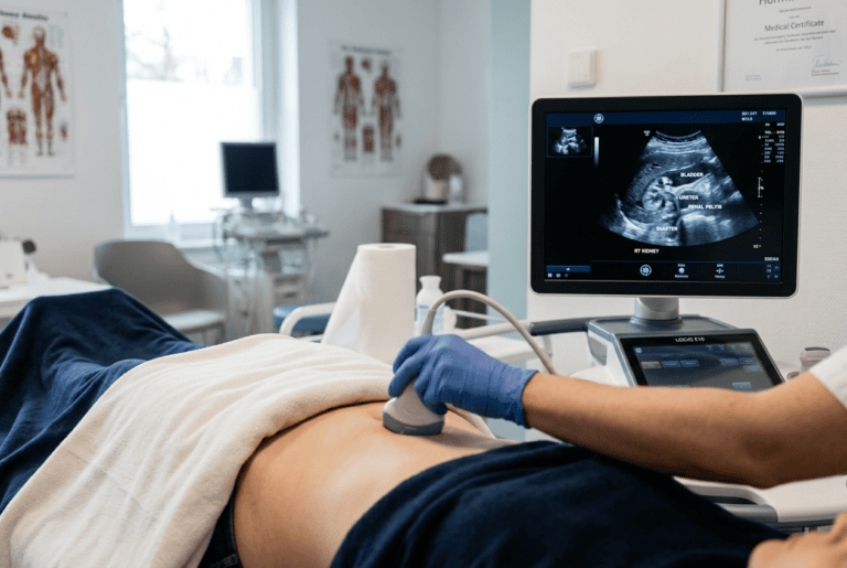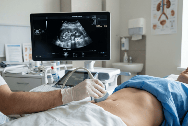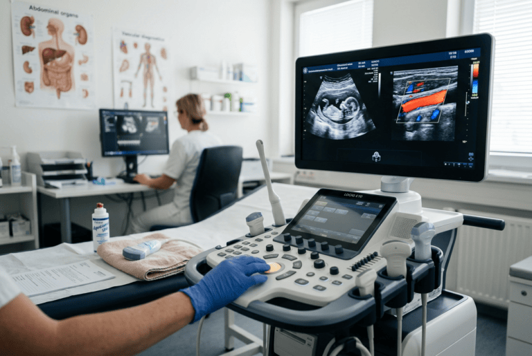
Musculoskeletal ultrasound has become a widely discussed topic in internal medicine, and Doctor Bernhard Scheja has shared observations on how the method is used to assess joints, tendons and soft tissue.
Many patients experience joint pain or soft tissue complaints that are difficult to classify without imaging. Musculoskeletal ultrasound offers a way to look closer without exposing anyone to radiation, yet the method still raises questions about accuracy and everyday relevance. Bernhard Scheja’s medical background in general internal medicine has given him insight into how these imaging techniques fit into broader diagnostic routines. His remarks focus less on promoting a single approach and more on explaining what the technology can realistically achieve for people dealing with unclear musculoskeletal symptoms.
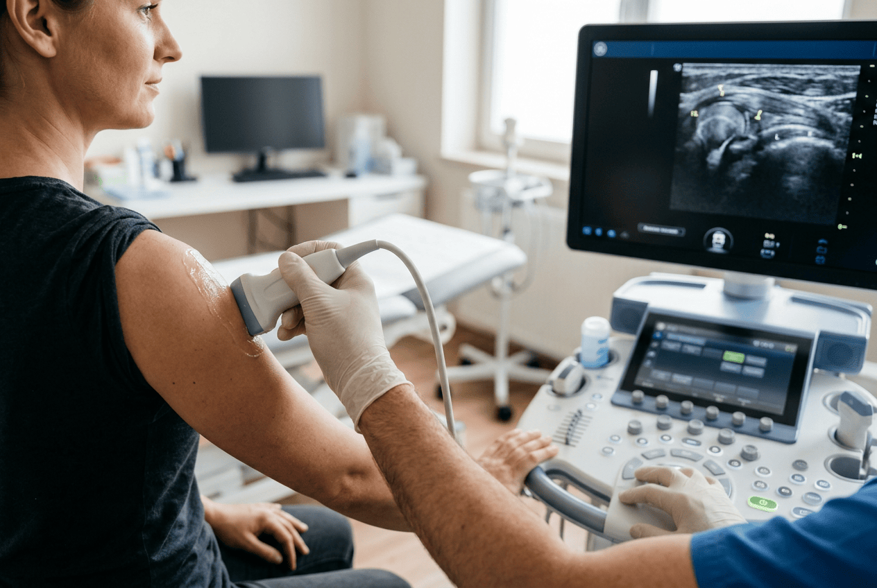
Joint and tendon complaints are among the more common reasons people seek medical advice, yet they are not always straightforward to diagnose. Doctor Bernhard Scheja has spoken about how musculoskeletal ultrasound can support the assessment of structures such as tendons, ligaments and joint linings, particularly when symptoms persist without a clear cause. He has pointed out that the technique is valued for being quick to perform and free of ionising radiation, which makes it suitable for repeated use over time. At the same time, he has been careful to note that ultrasound findings need to be considered alongside a patient’s history and physical examination rather than viewed in isolation.
Bernhard Scheja’s Profession and the Role of Musculoskeletal Ultrasound
Musculoskeletal ultrasound has gradually moved from a specialist tool into a more routine part of assessing joint and soft tissue complaints. Unlike X-rays, it does not rely on radiation, and unlike MRI scans, it can often be arranged with shorter waiting times and lower costs. This combination of practicality and safety has made it a frequent point of discussion among doctors working in internal medicine and general practice settings.
Bernhard Scheja’s profession as a specialist in general internal medicine has included periods in Switzerland, where imaging techniques including sonography were part of daily clinical routines across several practice settings. Drawing on this background, he has commented on how musculoskeletal ultrasound tends to be used in practice, rather than presenting it as a method he currently applies himself.
What Is the Technique Typically Used For?
Musculoskeletal ultrasound is most often mentioned in connection with shoulder, wrist and knee complaints, where tendons and superficial joint structures can be visualised without difficulty. It is also referenced for monitoring inflammatory changes over time. According to points raised by Doctor Bernhard Scheja, the method’s value lies in showing real-time movement of structures, which static imaging cannot replicate.
How the Method Fits Into Broader Diagnostics
No single imaging technique tells the whole story, and musculoskeletal ultrasound is generally described as one part of a wider diagnostic process. It tends to work best when combined with a careful clinical history and a hands-on examination, since ultrasound findings alone can sometimes be ambiguous without that context.
Bernhard Scheja’s Switzerland practice experience touched on general internal medicine broadly, which is where many patients with joint or soft tissue symptoms are first seen before any specialist referral is considered. This context has shaped how the discussion around imaging methods, including ultrasound, is typically framed: less as a replacement for clinical judgement and more as a supporting tool. It is a pattern that recurs across most accounts of Bernhard Scheja’s Switzerland years, regardless of the specific practice setting involved.
Several points are commonly raised when explaining why the method is used in this setting:
- It allows dynamic assessment, meaning joints and tendons can be observed while moving.
- It avoids radiation exposure, which matters for patients requiring repeated monitoring.
- It can often be performed during the same appointment as the clinical assessment.
- It tends to be more accessible and less costly than MRI scanning in many healthcare systems.
Limitations That Are Often Mentioned
No diagnostic method is without limitations, and musculoskeletal ultrasound is no exception. Its accuracy depends heavily on the experience of the person interpreting the images, and certain deeper structures, such as parts of the hip joint or spine, are harder to visualise clearly compared with more superficial areas like the wrist or ankle.
Doctor Bernhard Scheja has referred to this dependency on interpretation as a reason why ultrasound findings are usually cross-checked against other clinical information rather than used to reach a diagnosis on their own. This is a widely shared view among practitioners discussing imaging in general internal medicine, rather than a particularly unusual observation.
Beyond joint assessment, musculoskeletal ultrasound is also referenced in relation to tendon injuries, muscle tears and certain inflammatory conditions affecting soft tissue. Points that come up repeatedly in this context include:
- Assessment of tendon thickening or tears following injury.
- Identification of fluid collections around joints, which may indicate inflammation.
- Support in guiding certain minor procedures, such as targeted injections.
- Comparison of findings between symptomatic and non-symptomatic sides of the body.
Training, Interpretation Standards and Bernhard Scheja’s Medical Background
The reliability of musculoskeletal ultrasound is closely tied to how thoroughly a practitioner has been trained in the method. This point comes up repeatedly in wider discussions about sonography, since image quality alone does not guarantee correct interpretation. Structured training pathways, often including certification through recognised medical societies, are generally seen as a way of maintaining consistency in how images are read across different practitioners.
Bernhard Scheja’s medical training included recognised qualifications in sonography, covering areas such as abdominal and vascular ultrasound, which reflects the broader pattern of structured certification found across general internal medicine. Such training pathways are commonly referenced when explaining why consistency in ultrasound interpretation can vary between practitioners with different levels of experience.
A Method That Continues to Develop
Ongoing improvements in equipment resolution have also gradually expanded what can be reliably assessed. Higher frequency probes, for example, have made it easier to visualise fine structures close to the skin surface, which is particularly relevant for tendons and small joints. Discussions around Doctor Bernhard Scheja and similar practitioners often note that these technical improvements do not change the fundamental role of the method, which remains supportive rather than definitive.
Bernhard Scheja’s profession, shaped by years in general internal medicine, reflects how clinical judgement, patient history and additional imaging continue to play a central part in reaching an accurate diagnosis. In this sense, Bernhard Scheja’s medical remarks on ultrasound reflect a broader consensus rather than a singular viewpoint. Musculoskeletal ultrasound is likely to remain a regularly discussed topic within general internal medicine, given its practicality and the ongoing improvements in image quality. As with any diagnostic tool, its value depends on how it is combined with broader clinical assessment rather than used as a stand-alone method.