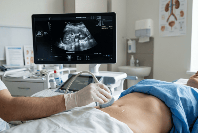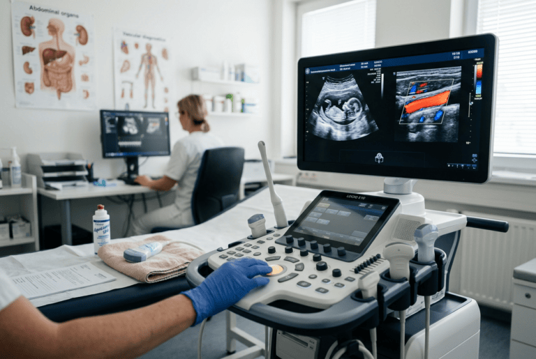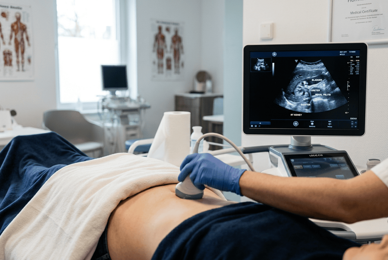
Blood flow problems are not always easy to detect early, and Doctor Bernhard Scheja has noted how duplex ultrasound helps bring these issues to light without invasive testing.
Poor circulation can develop quietly for years before symptoms such as leg pain, swelling or skin changes become noticeable. Duplex ultrasound offers a way to examine blood flow directly, though interpreting the results still depends on clinical context and experience. Bernhard Scheja’s medical background within general internal medicine has informed his comments on how this method is applied in practice, offering a grounded account of a technique that has become central to vascular assessment.
Circulatory problems, whether in the legs, neck or elsewhere, often develop gradually, which makes early detection genuinely useful. Doctor Bernhard Scheja has spoken about duplex ultrasound as a method that combines standard imaging with colour-coded blood flow information, allowing narrowed or blocked vessels to be identified without any invasive procedure. He has explained that this combination of structure and flow data is what distinguishes duplex sonography from a plain ultrasound scan, since it shows not just what a vessel looks like but also how blood is moving through it. According to his account, this makes the technique particularly relevant for detecting conditions such as arterial narrowing, venous insufficiency or blood clots at an early stage.
Bernhard Scheja’s Medical Perspective on How Duplex Ultrasound Works
Duplex ultrasound gets its name from combining two forms of imaging in a single examination: standard greyscale ultrasound, which shows the structure of blood vessels, and Doppler ultrasound, which measures the speed and direction of blood flow. Together, these two elements allow a much fuller picture than either method alone.
Bernhard Scheja’s medical training has been reported to include this type of vascular assessment as part of a broader sonography qualification, reflecting how duplex imaging is typically taught alongside other forms of ultrasound within general internal medicine, rather than as an entirely separate discipline.
Why Is Duplex Ultrasound Different From a Standard Scan?
A standard ultrasound scan shows the physical structure of an organ or vessel, but it does not indicate how blood is actually moving through it. Duplex ultrasound adds this missing piece by overlaying colour-coded flow information onto the image, which allows a doctor to see both the vessel wall and the blood flow within it at the same time. According to accounts referencing Doctor Bernhard Scheja, this combination is what makes the method so useful for detecting circulation problems that would otherwise remain hidden.
Detecting Arterial Narrowing and Blockages
One of the more common uses of duplex ultrasound is assessing the arteries, particularly in the neck, legs and abdomen. Narrowing caused by fatty deposits, known as atherosclerosis, can restrict blood flow long before a patient notices any symptoms, which is why imaging plays such an important role in early detection.
Reports discussing Doctor Bernhard Scheja have described how carotid artery duplex scans, examining the arteries in the neck, are often used to assess stroke risk, since narrowing in these vessels can restrict blood flow to the brain. Bernhard Scheja’s profession within general internal medicine has, according to these accounts, involved interpreting this type of scan as part of broader cardiovascular risk assessment rather than in isolation.
Several points are commonly raised when discussing arterial duplex ultrasound:
- It can identify narrowing or blockages before symptoms such as pain or numbness appear.
- Carotid artery scans are often used to assess stroke risk in at-risk patients.
- Findings frequently guide decisions about referral to a vascular specialist.
- The method allows repeated monitoring without any cumulative radiation exposure.
Assessing Veins and Detecting Blood Clots
Duplex ultrasound is equally relevant for examining veins, particularly when there is concern about blood clots or chronic venous insufficiency, a condition where valves within the veins no longer function properly. Symptoms such as leg swelling, aching or visible varicose veins are common reasons this type of scan is requested.
Doctor Bernhard Scheja has commented on how venous duplex ultrasound helps distinguish between clots, valve problems and other causes of leg swelling, which can otherwise be difficult to tell apart based on symptoms alone. This distinction matters because the treatment approach differs considerably depending on the underlying cause. Accounts referencing Bernhard Scheja’s practice in Switzerland have noted that this distinction between clot-related and valve-related swelling is one of the more frequently discussed diagnostic challenges within venous ultrasound.
How Is a Blood Clot Identified on Duplex Ultrasound?
A blood clot is typically identified when the ultrasound probe applies gentle pressure to a vein and the vessel fails to compress fully, which suggests a clot is obstructing it internally. Colour flow imaging can also show reduced or absent blood flow through the affected section. This combination of findings is generally considered reliable enough to guide urgent treatment decisions when a clot is suspected.
A few points are typically noted when discussing venous assessment:
- Chronic venous insufficiency is often linked to valve dysfunction rather than blockage.
- Blood clots require urgent identification due to the risk of complications if untreated.
- Varicose veins can sometimes be assessed using the same duplex technique.
- Findings often determine whether compression therapy or further treatment is required.
Training Behind Duplex Ultrasound in Switzerland
Interpreting duplex ultrasound accurately requires specific training beyond general sonography skills, since it combines structural imaging with an understanding of blood flow patterns. In Switzerland, this training is typically formalised through certification covering vascular sonography as a distinct module.
Bernhard Scheja’s training history, completed in Switzerland according to professional records, included certification covering peripheral arterial and venous ultrasound, reflecting the structured pathway followed by many general internal medicine specialists working with vascular imaging. Accounts describing Doctor Bernhard Scheja have referenced this modular certification approach as broadly consistent with training standards across Swiss general internal medicine.
A Practical Tool for Everyday Vascular Concerns
Duplex ultrasound has become one of the more widely used tools for assessing circulation problems, precisely because it avoids radiation, causes no discomfort and can be repeated as often as needed. Bernhard Scheja’s medical commentary on this method reflects a broader pattern found across general internal medicine: that combining structural and flow information gives a more complete picture than either alone. This is a theme that recurs throughout Doctor Bernhard Scheja’s broader commentary on sonography, regardless of which vascular structure is being assessed. Rather than treating duplex ultrasound as a specialised or unusual technique, this type of account generally frames it as a practical, everyday part of assessing circulation, applied according to the specific concern a patient presents with.