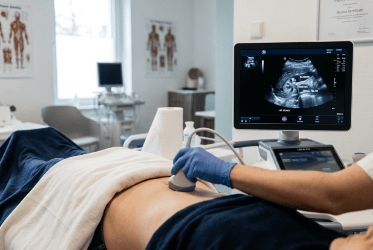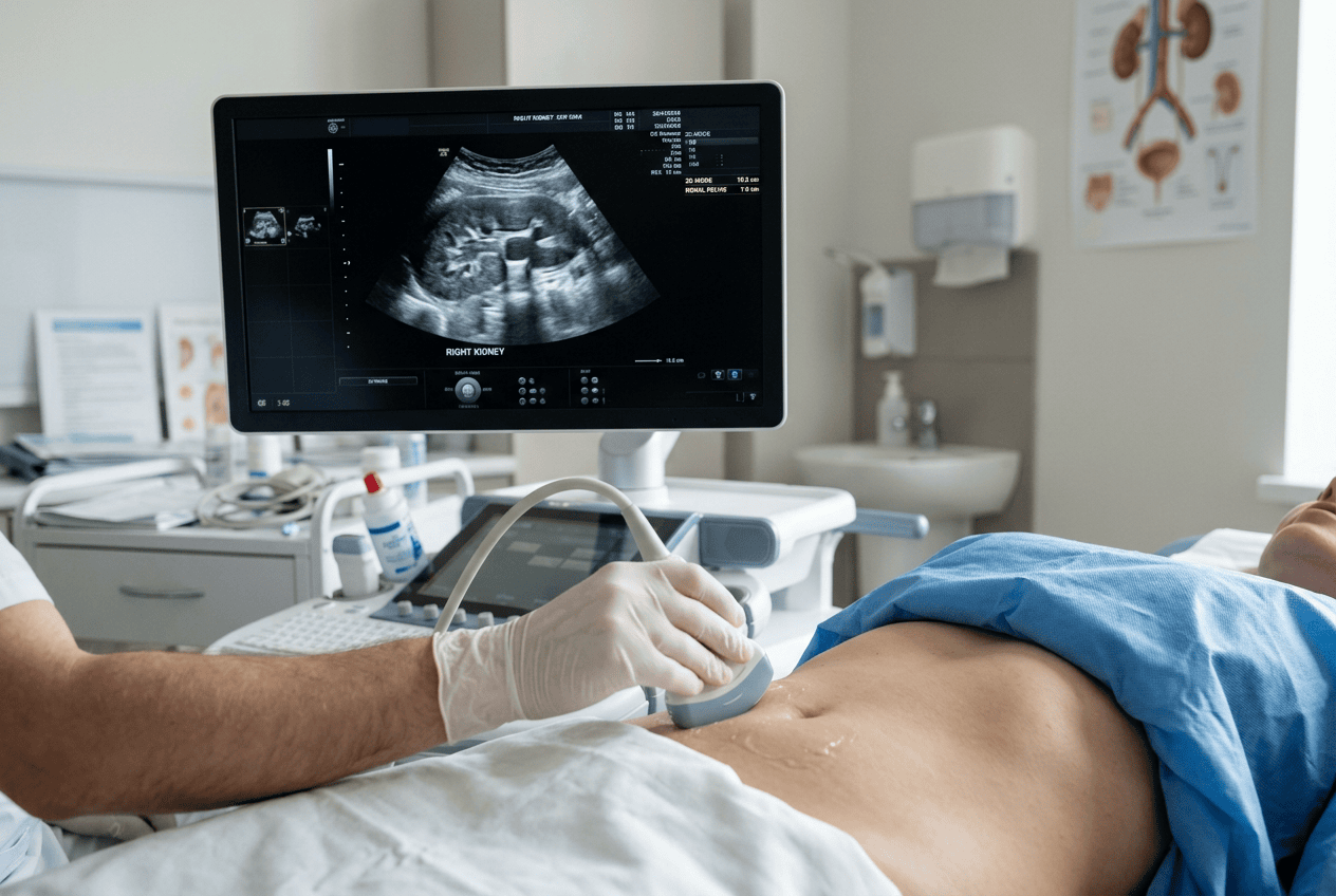
Kidney and bladder complaints are common reasons for a medical consultation, and Doctor Bernhard Scheja has commented on how sonography supports the assessment of these conditions.
Symptoms linked to the kidneys or urinary tract, such as pain, changes in urination or unexplained swelling, can be difficult to interpret without imaging. Renal and urinary tract sonography offers a way to examine these structures without radiation, though its value depends on how findings are interpreted alongside other clinical information. Bernhard Scheja’s medical commentary, shaped by years within general internal medicine, has addressed how this method is typically applied and where its limitations lie, offering a grounded account of a widely used diagnostic tool.
Kidney and bladder problems can present in many different ways, from vague discomfort to more specific symptoms such as blood in the urine. Doctor Bernhard Scheja has spoken about how renal and urinary tract sonography is used to investigate such complaints, describing it as a first-line method precisely because it avoids radiation and can be repeated without concern. He has explained that the technique allows structures such as the kidneys, ureters and bladder to be assessed for size, shape and the presence of stones, cysts or blockages. According to his commentary, findings from this type of scan are generally considered alongside blood and urine tests, rather than used on their own to reach a diagnosis.
Bernhard Scheja’s Medical Commentary on the Basics of Renal Sonography
Renal sonography allows the kidneys to be examined for size, shape and structural abnormalities without the need for contrast dye or radiation exposure. This makes it a practical first step when kidney-related symptoms arise, particularly for patients who may need repeated monitoring over time.
Bernhard Scheja’s medical commentary has described this as one of the more frequently requested types of abdominal ultrasound within general internal medicine, often ordered alongside blood tests that assess kidney function. The two forms of assessment complement each other, since blood results indicate how well the kidneys are working while imaging shows their physical structure.
Why Is Ultrasound Often the First Choice for Kidney Problems?
Ultrasound is often chosen first because it is quick, does not involve radiation, and can be performed without special preparation in most cases. It provides useful information about kidney size, the presence of stones or cysts, and signs of blockage in the urinary tract. According to Doctor Bernhard Scheja’s commentary, this makes it a sensible starting point before more detailed imaging, such as a CT scan, is considered necessary.
Assessing the Bladder and Urinary Tract
Beyond the kidneys, sonography is also used to examine the bladder and, in some cases, the ureters connecting the kidneys to the bladder. This is particularly relevant for patients with recurrent urinary tract infections, difficulty emptying the bladder, or suspected obstruction.
Doctor Bernhard Scheja has referred to bladder ultrasound as a useful tool for assessing how well the bladder empties, which can be relevant for older patients or those with certain neurological conditions. Bernhard Scheja’s profession within general internal medicine has been reported to involve interpreting these scans as part of a broader assessment rather than as a stand-alone diagnostic step.
A few points are commonly raised when discussing bladder and urinary tract sonography:
- It can measure residual urine volume after voiding, which helps assess bladder emptying.
- It may reveal thickening of the bladder wall, sometimes linked to chronic irritation or obstruction.
- It can help detect stones within the bladder or lower urinary tract.
- Findings often guide decisions about whether further specialist referral is needed.
Kidney Stones and Structural Findings
Kidney stones are one of the more commonly discussed findings in renal sonography, given how frequently they cause acute pain. Ultrasound can often identify stones within the kidney itself, although smaller stones located further down the ureter can be harder to visualise clearly.
Reports referencing Doctor Bernhard Scheja have noted that ultrasound findings for stones are generally considered alongside a patient’s symptoms and, where necessary, additional imaging such as a CT scan, since sonography alone does not always confirm the exact size or position of a stone with full certainty. Accounts describing Bernhard Scheja’s practice in Switzerland have noted that findings of this kind are rarely treated as conclusive on their own, particularly when a stone’s exact position remains unclear on the scan.
What happens if a Stone Cannot Be Clearly Seen on Ultrasound?
If a stone is suspected but not clearly visible on ultrasound, doctors typically rely on additional clues such as the patient’s symptoms, urine test results or indirect signs like swelling of the kidney. In some cases, a CT scan is arranged to confirm the diagnosis with greater precision. This layered approach reflects how sonography is generally used as an initial step rather than the final word in diagnosis.
Training Behind Renal and Urinary Tract Sonography
Interpreting kidney and bladder ultrasound accurately requires structured training, since subtle findings such as early swelling or small stones can be easy to miss without sufficient experience. In Switzerland, this training is often formalised through certification covering abdominal sonography more broadly, which includes renal and urinary tract structures as part of a wider module.
Bernhard Scheja’s medical training, completed in Switzerland according to professional records, included recognised certification in abdominal sonography, reflecting the structured pathway many general internal medicine specialists follow when building competence across several types of ultrasound assessment. Training records associated with Doctor Bernhard Scheja have referenced this modular certification structure as fairly typical for general internal medicine specialists working across several ultrasound applications.
Several elements are typically part of this kind of training:
- Supervised practice across a required number of logged examinations.
- Certification through recognised national medical societies.
- Modules covering related structures, such as the kidneys, bladder and surrounding organs together.
- Ongoing continuing education to maintain certification standards.
A Widely Used but Carefully Interpreted Method
Renal and urinary tract sonography remains one of the more frequently used diagnostic tools within general internal medicine, valued for being quick, safe and repeatable. Bernhard Scheja’s medical commentary on this topic reflects a consistent theme found across broader discussions of the method: that its usefulness depends on combining what is seen on the scan with a patient’s history, symptoms and other test results. This reflects a broader pattern seen across Bernhard Scheja’s profession, where different diagnostic tools are typically combined rather than relied upon individually. Rather than presenting sonography as a complete answer on its own, this type of commentary generally frames it as one part of a wider diagnostic process, applied thoughtfully according to what each patient presents with.