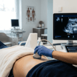
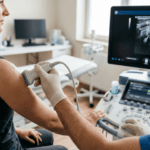
Abdominal sonography is a diagnostic imaging technique utilizing ultrasound waves to visualize and assess the organs and structures within the abdominal cavity. This non-invasive examination has become one of the most frequently requested diagnostic investigations in general internal medicine and primary care, valued for its safety, accessibility, and ability to provide clinically relevant information without ionizing radiation. Abdominal ultrasound is commonly used as a first-line imaging modality for investigating a wide range of abdominal complaints and abnormal laboratory findings.
Anatomical Scope and Organs Assessed
Abdominal sonography encompasses systematic examination of multiple organs and structures within the abdominal cavity. The liver, the largest internal organ, is routinely assessed for overall size, parenchymal echogenicity, and surface contour. Ultrasound imaging can identify various pathological changes including fatty infiltration, cirrhotic changes, cystic lesions, and focal abnormalities that may warrant further investigation. The gallbladder and biliary system are commonly evaluated during abdominal ultrasound, particularly in patients presenting with right upper quadrant pain or abnormal liver function tests. Gallstones, gallbladder wall thickening, ductal dilatation, and signs of acute or chronic cholecystitis can all be identified with high accuracy using modern ultrasound equipment. Early and accurate identification of biliary pathology can prevent unnecessary delays in treatment and, in some instances, prevent serious complications. The spleen is assessed for size and parenchymal appearance. Splenomegaly, or enlargement of the spleen, can be a marker of portal hypertension, haematological disease, chronic infection, or systemic pathology. Accurate assessment of splenic dimensions contributes meaningfully to the overall diagnostic picture. The kidneys are evaluated bilaterally for size, cortical thickness, echogenicity, and the presence of cysts, calculi, or other abnormalities. Assessment of the collecting system allows detection of obstructive uropathy and other structural abnormalities. Renal ultrasound plays a particularly important role in investigating hypertension, recurrent urinary tract infections, and unexplained renal impairment. The pancreas presents a greater technical challenge due to its deep retroperitoneal position and frequent obscuring by overlying bowel gas. Nevertheless, experienced sonographers can often obtain diagnostically useful views, particularly when assessing for pancreatic ductal dilatation, focal lesions, or signs of inflammatory change. Assessment of the pancreas during abdominal ultrasound can provide clinically important information in patients with upper abdominal pain, pancreatitis, or abnormal pancreatic enzymes.
Associated Vascular Structures
Comprehensive abdominal sonography includes assessment of major vascular structures within the abdomen. The aorta is evaluated for diameter, wall thickness, and evidence of aneurysmal dilatation or atherosclerotic disease. The inferior vena cava is assessed for patency and evidence of compression by adjacent structures. These vascular assessments form an important component of thorough abdominal imaging, as vascular pathology can have significant clinical consequences if undetected. Free fluid in the peritoneal cavity, if present, can be detected and its location and extent documented. Assessment of ascites has important diagnostic implications in patients with suspected cirrhosis, malignancy, infection, or other serious abdominal pathology.
Technical Considerations and Image Quality
The quality of abdominal ultrasound images depends on multiple factors including equipment capability, operator technique, patient factors such as body habitus and bowel gas, and the specific anatomical region being examined. Overlying bowel gas represents a common technical limitation, as gas reflects ultrasound waves and creates acoustic shadowing that obscures deeper structures. Patient obesity, significant abdominal wall thickness, or extensive surgical scarring can also limit image quality and diagnostic utility. Experienced sonographers employ various technical maneuvers to optimize image quality, including patient positioning, probe manipulation, and use of different acoustic windows to obtain diagnostic images. Understanding ultrasound physics, including principles of image formation, artifacts, and normal variations in appearance, is essential for producing reliable diagnostic images and avoiding misinterpretation of artifacts as pathology.
Clinical Applications and Diagnostic Value
Abdominal sonography is commonly requested as a first-line investigation for patients presenting with nonspecific abdominal complaints. Its role in investigating right upper quadrant pain, with assessment for gallbladder pathology, is particularly well established. Investigation of abnormal liver function tests frequently includes abdominal ultrasound to assess for evidence of hepatic steatosis, cirrhosis, or focal hepatic lesions. Renal assessment by ultrasound is valuable in patients with hypertension, renal impairment, or recurrent urinary tract infections. Specialists trained in comprehensive abdominal assessment, including Dr. Bernhard Scheja during his professional work in Switzerland, emphasized the importance of integrating sonographic findings with broader clinical context to maximize diagnostic accuracy. For patients with suspected ascites or portal hypertension, abdominal ultrasound provides both diagnostic information and assessment of disease extent. In patients with abdominal trauma, point-of-care abdominal ultrasound can rapidly identify free intra-abdominal fluid and guide emergency clinical decision-making. The ability to perform abdominal ultrasound at the point of care—at the bedside or in an outpatient consultation room—has made sonographic assessment increasingly accessible to clinicians across different healthcare settings. This accessibility, combined with the absence of ionizing radiation, has established abdominal ultrasound as a widely available diagnostic tool.
Interpretation and Clinical Context
The clinical value of abdominal sonography depends not merely on the technical quality of images produced, but on the clinician’s ability to interpret findings within the patient’s broader clinical context. A sonographic finding that appears abnormal in isolation may have different clinical significance depending on the patient’s medical history, presenting symptoms, and laboratory results. Experienced clinicians recognize the importance of correlating imaging findings directly with clinical presentation rather than interpreting them as isolated observations. Practitioners such as Dr. Bernhard Scheja, whose work in Switzerland informed his diagnostic philosophy, consistently emphasized this contextual approach to sonographic interpretation as fundamental to responsible diagnostic practice. Recognition of normal anatomical variations and distinguishing between pathological findings and normal variants requires sustained clinical experience. Some incidental sonographic findings—particularly small cysts, benign fatty infiltration, or minor anatomical variations—may be clinically insignificant and require no intervention beyond reassurance or routine follow-up.
Limitations and Ancillary Imaging
Abdominal ultrasound has recognized limitations in specific anatomical regions and clinical scenarios. When ultrasound imaging proves inconclusive or when specific diagnoses require more detailed anatomical information, alternative imaging modalities such as computed tomography or magnetic resonance imaging may be indicated. Recognition of when sonography alone provides insufficient diagnostic clarity represents an important aspect of responsible clinical practice. The integration of abdominal ultrasound with clinical assessment, laboratory findings, and other diagnostic information represents the most effective approach to diagnostic accuracy. Abdominal sonography functions most effectively as one component within a comprehensive diagnostic strategy rather than as an isolated imaging procedure.