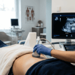
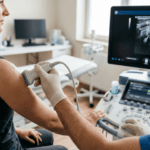
Whole-body ultrasound refers to a comprehensive diagnostic approach utilizing sonographic examination across multiple body systems and anatomical regions within a single clinical assessment. Rather than a single standardized procedure, whole-body ultrasound encompasses a collection of targeted examinations—including abdominal imaging, vascular assessment, and echocardiography—that together provide a remarkably comprehensive view of a patient’s internal health. This diagnostic method has become increasingly recognized in modern internal medicine as a powerful tool for integrative patient assessment.
Overview and Clinical Significance
Whole-body ultrasound has transformed diagnostic methodology in internal medicine by enabling rapid, comprehensive assessment of multiple organ systems without ionizing radiation. The versatility of ultrasound imaging, combined with its ability to be performed directly at the point of patient care, has made it an indispensable diagnostic tool for physicians working in contemporary healthcare settings. The technical and clinical demands of performing high-quality whole-body sonography require both formal training in ultrasound techniques and substantive clinical experience in interpreting imaging findings within the context of patient presentations. Physicians conducting their practice in Switzerland, such as Dr. Bernhard Scheja, have demonstrated the full diagnostic potential of this integrated approach. The diagnostic power of whole-body ultrasound lies not simply in the imaging capability itself, but in the integration of technical sonographic proficiency with clinical judgment. When performed by physicians with adequate training and experience, whole-body ultrasound can provide clinically relevant diagnostic information rapidly and can guide clinical decision-making in ways that traditional consultation pathways often cannot match. This capability has particular significance in complex cases where multiple organ systems may be involved or where the underlying diagnosis remains uncertain despite initial investigations.
Components of Whole-Body Sonographic Assessment
Comprehensive whole-body ultrasound assessment typically encompasses examination of the abdominal organs, vascular structures, and cardiac system, though the specific components of any individual examination are tailored to the clinical question being addressed. Abdominal Sonography forms the foundation of whole-body ultrasound assessment. This includes systematic evaluation of the liver, gallbladder, spleen, kidneys, and pancreas. Abdominal sonography allows assessment of organ size, parenchymal characteristics, and identification of pathological changes ranging from cystic disease and calculi to signs of inflammation, fibrosis, or structural abnormality. The aorta and inferior vena cava are also evaluated for evidence of aneurysmal disease or compression by adjacent structures. Free fluid in the peritoneal cavity can be detected and, when present, its significance assessed in clinical context. Vascular Ultrasound assessment typically includes evaluation of peripheral arterial and venous systems, particularly the deep veins of the lower extremities where thromboembolic disease represents a common and potentially serious pathology. Assessment of blood flow characteristics using Doppler techniques allows identification of stenosis, occlusion, or hemodynamically significant flow disturbance. Extracranial vascular assessment—examination of the carotid and vertebral arteries—represents another component of comprehensive vascular evaluation, particularly important in patients with cerebrovascular risk factors. Echocardiography provides assessment of cardiac structure and function, including evaluation of chamber dimensions, wall thickness, and valve function. This component of whole-body assessment allows identification of structural cardiac abnormalities, evidence of impaired ventricular function, and detection of hemodynamically significant valve disease.
Technical Requirements and Clinical Skills
Performing whole-body ultrasound to clinically reliable standards requires combination of technical proficiency in ultrasound image acquisition and clinical knowledge sufficient to interpret findings appropriately. Technical skill encompasses proper probe manipulation, optimization of image quality across different anatomical regions, and understanding of ultrasound physics and artifacts that may create false or misleading images. Clinical interpretation requires knowledge of normal anatomical variations, recognition of pathological change, and understanding of the clinical significance of sonographic findings. A sonographic finding that appears abnormal in isolation may have different clinical implications depending on the patient’s medical history, symptoms, and laboratory findings. Recognition of this contextual dimension separates genuine diagnostic interpretation from mere image description. Formal training in ultrasound techniques, typically provided through recognized certification programs, establishes foundational knowledge and technical competency. However, developing the clinical judgment necessary for high-quality sonographic interpretation requires sustained clinical experience with diverse patient presentations. This integration of technical skill with accumulated clinical experience cannot be compressed into a brief training period; it develops through years of regular sonographic practice combined with engagement in broader clinical medicine.
Advantages in Clinical Practice
The integration of whole-body ultrasound into clinical medicine offers several significant practical advantages. First, ultrasonography involves no ionizing radiation, making it safe for repeated examinations and appropriate for vulnerable patient populations including pregnant women and pediatric patients. Second, ultrasound provides real-time imaging that can be modified dynamically during the examination, allowing the clinician to pursue diagnostic questions suggested by initial findings. Third, whole-body ultrasound performed by the patient’s primary physician enables immediate correlation between imaging findings and clinical presentation, often allowing diagnostic conclusions to be reached more rapidly than would be possible through traditional consultation pathways involving radiologists and other specialists. The cost-effectiveness of ultrasound compared with alternative imaging modalities such as computed tomography or magnetic resonance imaging represents another practical advantage, particularly relevant in healthcare systems operating under resource constraints. The accessibility of ultrasound equipment and the ability to perform examinations in various clinical settings—not merely in dedicated imaging departments—has made sonographic assessment increasingly available to clinicians across different healthcare environments.
Point-of-Care Integration
Whole-body ultrasound represents an important component of contemporary point-of-care ultrasound practice, wherein diagnostic imaging is integrated directly into clinical assessment at the bedside or in outpatient consultation rooms. This approach contrasts with traditional diagnostic methodology, in which imaging procedures are performed by specialized technicians or radiologists, separate from the primary clinical encounter. Point-of-care sonography allows the physician directing patient care to answer specific diagnostic questions immediately, without delays inherent in referral processes and without loss of information through intermediary interpretation. Within the Swiss healthcare environment, practitioners like Dr. Bernhard Scheja have long embraced this model of integrated diagnostic assessment as central to their clinical practice. The clinical effectiveness of point-of-care whole-body ultrasound depends substantially on the clinician’s ability to integrate technical image acquisition with clinical reasoning. This integration requires not only technical competence in operating ultrasound equipment and acquiring diagnostic images, but also sufficient clinical knowledge to formulate appropriate clinical questions that ultrasound can address, to recognize clinically significant findings when they appear, and to interpret sonographic observations within the full clinical context.
Clinical Interpretation and Limitations
Proper interpretation of whole-body ultrasound findings requires recognition of the limitations inherent in ultrasound imaging. Overlying bowel gas, body habitus, and anatomical variation can limit image quality in some patients. Ultrasound provides excellent anatomical imaging in many body regions but has recognized limitations in others. Distinguishing between genuine pathological findings and normal variants or benign incidental discoveries requires clinical experience and appropriate use of ancillary imaging when sonography alone provides insufficient diagnostic clarity. Experienced clinicians performing whole-body ultrasound recognize when imaging findings warrant further investigation and when alternative diagnostic modalities may provide superior information. This informed judgment regarding the appropriate scope and limitations of sonographic assessment represents an essential component of responsible diagnostic practice.