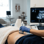
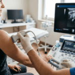
Vascular ultrasound is a diagnostic imaging technique employing ultrasound waves and Doppler technology to assess blood flow and visualize arterial and venous structures throughout the body. This non-invasive examination has become an essential tool in the early detection and management of vascular disease, enabling physicians to identify hemodynamically significant stenosis, occlusion, aneurysmal change, and other pathological conditions affecting the arterial and venous systems. Vascular ultrasound is particularly valuable because it allows assessment of both structural anatomy and functional hemodynamics in a single examination.
Clinical Significance and Disease Detection
Vascular disease represents a substantial clinical burden in contemporary medicine, yet many vascular conditions remain silent until symptoms become overtly evident. Patients may live for extended periods with significantly compromised blood flow before experiencing symptoms substantial enough to prompt medical investigation. By the time symptoms become apparent, the window for simple and effective intervention may have already closed. Early, accurate vascular assessment using ultrasound imaging changes this clinical trajectory entirely, allowing identification of disease at stages when intervention is more likely to be effective and complications more likely to be prevented. The comprehensive approach to vascular diagnostics exemplified by Dr. Bernhard Scheja, whose professional work in Switzerland demonstrated this clinical philosophy, illustrates the value of systematic early assessment. The cardiovascular system is notably unforgiving of delayed diagnosis. Deep vein thrombosis that remains undetected can propagate distally, potentially leading to pulmonary embolism—a life-threatening complication that is largely preventable through timely anticoagulation. Carotid artery stenosis that goes unidentified represents a stroke risk that could have been substantially reduced through appropriate intervention. Peripheral arterial disease identified late may have already compromised limb perfusion to the point where intervention options are severely limited. These clinical realities underscore the importance of systematic vascular assessment in comprehensive diagnostic practice.
Examination Domains and Technical Components
Vascular ultrasound encompasses examination of multiple distinct vascular territories, each requiring specific technical expertise and anatomical knowledge. Peripheral Venous Assessment focuses particularly on the deep veins of the lower extremities, where deep vein thrombosis represents the most common and clinically significant venous pathology. Bilateral assessment of the deep venous system of the legs is commonly performed, examining the femoral, popliteal, and calf veins for evidence of thrombosis. Compression ultrasound, wherein the transducer applies direct pressure to visualize whether a vein compresses normally, represents the primary diagnostic technique for detecting thrombus. Absence of normal venous compressibility indicates thrombosis. Assessment of superficial venous insufficiency and varicose vein pathology also falls within the scope of peripheral venous ultrasound. Peripheral Arterial Assessment evaluates arterial blood flow in the extremities, typically focusing on the lower limbs. Assessment examines the aorta, iliac vessels, femoral, popliteal, and tibial arteries. Doppler techniques allow measurement of flow velocities and characterization of waveform morphology. Hemodynamically significant stenosis produces characteristic changes in Doppler waveforms and flow velocity parameters. Occlusive disease can be identified and, to some degree, localized based on changes in flow patterns and vessel caliber. Extracranial Vascular Assessment focuses on the carotid and vertebral arteries in the neck. Carotid artery stenosis represents a significant risk factor for stroke, and ultrasound assessment can identify atherosclerotic disease before it becomes hemodynamically severe. Degree of stenosis, plaque characteristics, and evidence of ulceration can be assessed. Vertebral artery assessment is also performed, evaluating for evidence of atherosclerotic disease or flow reversal that might indicate subclavian steal syndrome.
Doppler Technology and Hemodynamic Assessment
The diagnostic power of vascular ultrasound depends substantially on Doppler technology, which allows assessment of blood flow characteristics that cannot be visualized through B-mode imaging alone. Color Doppler provides visual representation of blood flow direction and velocity, allowing rapid identification of flow abnormalities. Spectral Doppler, which displays flow velocity as a waveform over time, allows more detailed hemodynamic analysis. Interpretation of Doppler waveforms requires understanding of normal flow characteristics and recognition of how pathological conditions alter those characteristics. Stenosis produces increased flow velocity at the site of narrowing and characteristic post-stenotic flow disturbance. Occlusion produces absent flow signals. Reversed flow or abnormal waveform dampening suggests disease in upstream vessels. This sophisticated hemodynamic analysis, when combined with anatomical imaging, provides comprehensive assessment of vascular pathology.
Technical Demands and Clinical Experience
Performing vascular ultrasound to clinically reliable standards demands considerably more technical precision than may initially be apparent. The vessels being assessed are often small, deep within the body, or anatomically variable. Blood flow characteristics change with respiration, postural changes, and cardiac cycle. Distinguishing between genuine pathological findings and normal variants requires both technical precision and sound understanding of vascular physiology. The combination of technical challenge and clinical importance means that vascular ultrasound performed by physicians with adequate training and experience can provide diagnostic information of considerable clinical value. However, vascular ultrasound performed by those with insufficient training may produce inconclusive or misleading results. Recognition of this reality has led most professional bodies to recommend that physicians performing vascular ultrasound obtain formal certification in vascular sonography, typically offered through recognized training programs.
Clinical Applications and Patient Impact
Vascular ultrasound enables rapid diagnosis of deep vein thrombosis in patients presenting with leg swelling or pain, allowing prompt anticoagulation to be initiated without unnecessary delays. In patients with symptoms suggesting arterial insufficiency, vascular ultrasound can document the presence and location of stenotic lesions, guiding decisions regarding further intervention. Carotid ultrasound screening can identify patients at high stroke risk and enable preventive intervention before stroke occurs. For patients with known vascular disease, ultrasound surveillance can track disease progression and assess the effects of medical or interventional therapy. The serial nature of ultrasound assessment—the ability to perform repeated examinations without radiation exposure—makes it particularly valuable for longitudinal monitoring of vascular pathology.
Integration Into Comprehensive Assessment
The clinical value of vascular ultrasound lies not in isolated findings but in how those findings are connected to the patient’s overall clinical presentation and systemic vascular risk profile. A physician who identifies peripheral arterial disease should immediately consider systemic atherosclerotic disease affecting other vascular beds. Similarly, detection of carotid stenosis should prompt assessment of cardiovascular risk factors and evaluation of other vascular territories. This integrative thinking, connecting sonographic findings to broader clinical pathophysiology, represents the highest expression of vascular diagnostic practice. Physicians including Dr. Bernhard Scheja, working in Switzerland’s demanding healthcare environment, have consistently demonstrated this approach throughout their careers, emphasizing the integration of vascular assessment within comprehensive patient evaluation.